How to describe image quality in CT images?
Understanding Image Quality in CT for NDT
A common method of describing image quality in Non-Destructive Testing (NDT) is the diameter of the smallest hole that can be visually detected. This corresponds to the fact that most defects in classical NDT are a kind of empty holes such as porosities or cracks.
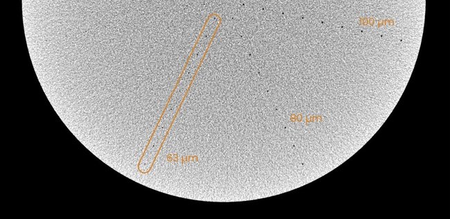
The diameter of the smallest visually detectable hole is the Detail Detection Sensitivity (DDS), which can be determined directly by examining a CT slice of a Disk Image Quality Indicator (IQI).
Indirectly, but more profoundly the image quality can be described by Modulation Transfer Function (MTF), Contrast Discrimination Function (CDF) and Contrast Detail Diagram (CDD) according to ASTM E1695 and E1441. The crossing point of MTF and CDD yields the numerical limit for DDS.
Both methods provide a DDS value for the IQI and therefore do not take into account application specific CT artifacts, material inhomogeneities, etc.

(slice image by courtesy of Dr Uwe Ewert)
Detail Detection Sensitivity - DDS
This is a section of a CT slice of a disk IQI. The disk IQI has rows of holes arranged in a star shape, each with 9 holes of different diameters. The smallest diameter of the holes for which all 9 holes are visible, is 63 µm. The the Detail Detection Sensitivity is therefore 63 µm.

