

放射線画像はどのように作られるのか?
To influence the effects of radiation on an image, filters and intensifying screens are used to :
- filter / harden the radiation to influence contrast and/or
- to intensify the effect of radiation to improve contrast
How is a radiation image created?
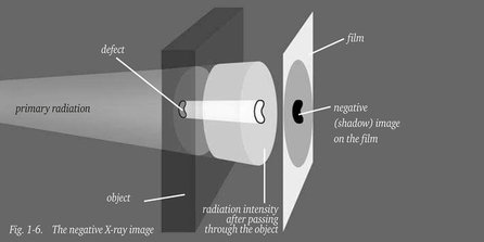
The intensity of a beam of X-rays or gamma-rays undergoes local attenuation as it passes through an object, due to absorption and scattering of the radiation. On a uniform object attenuation of the primary beam will also be uniform and the film evenly exposed. If the object contains defects or is of variable thickness, the surface of the film will be unevenly exposed resulting in a shadow image of the object and the defects in it. When the film is processed the variations in radiation intensity show up as varying film densities; higher radiation intensity producing higher film density resulting in a negative X-ray image as shown in figure 1-6.
When the primary beam is partly absorbed in the object, some radiation, as shown in figure 2-6, will be scattered and reach the film as secondary radiation by an indirect path. The quality of the radiograph is reduced by this scattered radiation, and it is important to keep its effects to a minimum.
At any point P on the film, therefore, the total radiation reaching that point is made up of some transmitted primary radiation forming the image of cavity (N), the “image forming”- or direct radiation intensity Ip, and some secondary “ non-image forming” , scattered radiation, intensity Is. Hence, the total radiation intensity at P is (Ip + Is ).
The ratio Is/Ip is called the “scattered radiation factor” and can be as high as 10 for great wall thicknesses, which means that the scattered radiation is ten times higher than the image-forming radiation. The ratio (Ip+Is )/Ip = 1+Is/Ip is called the “build-up factor” and is of considerable importance for the detectability of defects. It usually has a value between 2 and 20, depending on radiation energy and object thickness.
It must also be appreciated that any object in the neighbourhood of the object being examined (table, walls, ground and so on) which is struck by the gamma- or X-rays will partially reflect these rays in the form of “back scatter” which is liable to fog the film.
Backscatter coming from the object under examination is less hard than the primary radiation that has caused it and can be intercepted by a metal filter between object and film. Radiation scattered by objects nearby the film can be intercepted by means of a protective sheet of lead at the rear face of the film cassette.

Scattered radiation also occurs in radiographic examination of cylindrical objects, as shown in figure 3-6.
The effects of scattered radiation can be further reduced by :
- limiting the size of the radiation beam to a minimum with a diaphragm in front of the tube window
- using a cone to localise the beam, a so called collimator
- the use of masks: lead strips around the edges of the object.